Laser removal of a port-wine stain (nevus flammeus)
- Aug 17, 2025
- 3 min read

A flaming nevus (naevus flammeus, port-wine stain) is a congenital vascular malformation of the skin, appearing as a bright pink, red, or purple spot, most often located on the face, neck, or upper body.
This is not merely an aesthetic feature — a flaming nevus can significantly affect a patient’s quality of life, self-esteem, and psychological well-being, especially in children and adolescents.
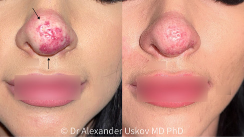
At the clinic of international expert Dr. Uskov, we use advanced laser technology to correct congenital vascular anomalies, employing all necessary internationally certified wavelengths. We aim for maximum lightening or even complete removal of the lesion, with a focus on safety, predictability, and an individualized approach.
Relevance of the issue
A flaming nevus occurs in approximately 0.3–0.5% of newborns. It can persist throughout life, and without treatment, may darken and thicken with age.
The most common locations include the face, particularly:
cheeks,
forehead,
eyelids,
neck and occipital area.
Although the lesion does not pose a medical threat, it can be:
cosmetically disturbing,
a source of psychological stress for children and adults,
a marker of systemic syndromes (e.g., Sturge–Weber syndrome — requiring differential diagnosis).
History of the condition
The term “port-wine stain” was introduced into medical practice in the 20th century due to the characteristic color of the lesion. Before the advent of lasers, such vascular formations were nearly impossible to treat. It was only in the 1980s, with the introduction of pulsed dye lasers (PDL), that a new era in the treatment of vascular malformations began.
Why is a laser necessary for treatment?
Selective action — the laser energy is absorbed by hemoglobin in the vessel without damaging the surrounding skin
Minimal trauma — rapid recovery with no scarring
High effectiveness — especially when treatment is started at an early age
No systemic effect — particularly important for children and adolescents
The treatment course
Consultation and diagnosis
During the first appointment, the doctor:
assesses the depth and density of the vascular network;
performs digital dermatoscopy;
uses a UV-light, infrared imaging, and, if necessary, ultrasound
Procedure
Duration: 5–15 minutes depending on the treated area.
Age: treatment is potentially possible from 1 year of age (or earlier if indicated), but the decision is made individually.
Anesthesia: usually not required; local anesthesia may be used if needed.
Eye protection: scleral shields are used for lesions near the eyes.


How many session do I need?
Average number of sessions for optimal results:
Children: 2–5 sessions at intervals of 6–12 weeks
Adults: 3–7 sessions, rarely more
The number of sessions depends on:
skin thickness and vessel depth;
age;
area of the lesion;
previous treatments (if any);
skin phototype.
Safety is our highest priority
Treating congenital vascular anomalies requires:
precise anatomical diagnostics;
risk assessment;
experience with children and sensitive areas (eyelids, forehead, nose).
At our clinic:
procedures are performed by doctors with qualifications in laser medicine;
advanced certified equipment is used
Post-treatment care
After the procedure, patients may experience:
redness, swelling, or bluish tint — usually resolve within 2–5 days;
occasionally, small crusts that fall off naturally.
Recommendations:
avoid UV exposure;
use sunscreens (SPF 50+);
avoid mechanical irritation of the skin;
follow the doctor’s individual care instructions.
Individual results
Every vascular lesion is unique. Effectiveness depends on:
the maturity of the vascular tissue;
individual skin characteristics;
skin type (Fitzpatrick scale);
systemic conditions (e.g., hormonal fluctuations).


A congenital port-wine stain is not the end of the world. Modern laser technologies, combined with medical expertise, allow for the delicate and safe reduction of the vascular lesion.