
Do laser treatments reduce the risk of skin cancer?
Scientific evidence as of 21 October 2026
Yes, studies have already shown this effect for some laser treatments: fewer new tumours developed in the treated skin. This is encouraging news for laser dermatology. Renewing sun-damaged skin may offer benefits that go beyond its appearance. [1, 2, 3, 4]
The research mainly concerns basal cell carcinoma and squamous cell carcinoma. Most studies have involved people with substantial sun damage or a previous history of skin cancer. Laser treatments have not yet been shown to reduce the risk of melanoma.
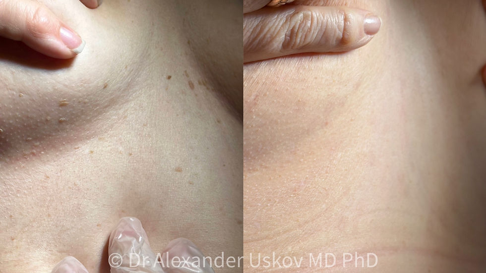
These photographs show examples of improvements in pigment spots, uneven skin tone and surface irregularities. The aesthetic changes are clearly visible, and the potential benefits of some laser methods may extend further: studies suggest that they could reduce the risk of new basal cell and squamous cell carcinomas in sun-damaged skin. [1, 2, 3, 4]
This is an additional, promising area of research. The photographs illustrate the visible results of procedures; any potential reduction in cancer risk is assessed separately, through long-term follow-up. The specific treatment is chosen after examining the skin and assessing whether it is appropriate.

Why might this happen?
The sun gradually changes our skin: pigment spots, wrinkles and an uneven surface develop. These changes are known as photoageing. At the same time, ultraviolet radiation can damage cells and increase the risk of skin cancer. Not all the effects of sun exposure are visible in the mirror. [5]

One sign of this damage is actinic keratosis. It usually looks like a small, rough, scaly patch. Some of these lesions can develop into squamous cell carcinoma over time, so it is important to have them assessed by a doctor. However, not every pigment spot or area of flaky skin is an actinic keratosis. [5]
A laser can remove some of the damaged tissue and trigger skin renewal. Fractional lasers are particularly interesting: they treat many tiny areas, leaving untouched skin between them. Studies suggest that this treatment also improves the function of cells involved in skin repair and the response to ultraviolet radiation. This is one possible mechanism behind a reduction in the risk of tumours. [12, 15]
Aesthetic dermatology also addresses individual raised skin lesions and surface irregularities. A doctor assesses their nature and whether removal is appropriate before treatment. These photographs do not identify the lesions shown as precancerous.

How did the idea develop?
As early as the 1990s, doctors began investigating whether laser resurfacing could reduce the number of actinic keratoses and new skin tumours. The first studies were small, and their results varied. More convincing evidence emerged in the 2000s: resurfacing severely sun-damaged skin reduced the number of keratoses, and some studies also found fewer new tumours. [6, 7, 8, 9, 10, 11]
Animal studies later showed that fractional laser treatment could also delay the development of tumours caused by ultraviolet radiation. Studies with long-term follow-up of patients were the next important step. [13, 14]

What did long-term follow-up show?
A study published in 2021 involved 48 people aged 60 or older with substantial sun damage on their arms. One arm received fractional laser treatment, while the other was left untreated. This allowed researchers to compare treated and untreated skin in the same person. [1, 18]
During up to three years of follow-up, 24 tumours were found in the untreated areas, compared with 2 in the treated areas. This count included the earliest forms of squamous cell carcinoma, confined to the skin’s surface layer. These figures refer to tumours, not patients: one person could develop more than one lesion. [1]
Some participants in the same group were followed for up to five and a half years. The follow-up study, published in 2026, continued to find fewer new tumours and keratoses in the treated skin. The beneficial effect of a single treatment was therefore observed over several years. [2]

What do we know about other lasers?
A 2023 study compared patients who had previously had skin cancer on the face. New tumours developed in approximately 21% of those who received nonablative fractional treatments — treatments that do not vaporise the skin’s surface — compared with 40% of those who did not. [3]
A similar association was found for pulsed dye laser treatment, which is usually used for vascular skin changes. In a study published online in 2025 and in a journal issue in 2026, new tumours were found in approximately 27% of patients who had received laser treatment and 54% of the comparison group. All participants had previously had skin cancer on the face. [4]
Both studies were based on reviews of medical records. They show an encouraging association, although the effect of the laser cannot yet be fully separated from other differences between the patients.

There is encouraging news about thulium lasers too. Studies, including one published in 2026, found reductions in actinic keratoses and signs of photoageing. Improvements were also visible under the microscope. These studies examined the condition of damaged skin; the incidence of skin cancer in subsequent years still needs to be assessed. [17, 19]

Researchers are also studying lasers in combination with photodynamic therapy, a treatment in which light activates a special medicine applied to the skin. In a 2026 study, this combination helped clear keratoses more reliably than photodynamic therapy without laser pretreatment. [20]

How strong is the evidence for a lower risk?
There is already direct clinical evidence: fewer new tumours developed in skin treated with a particular fractional laser procedure. Other studies support this line of research. The possibility of reducing skin cancer risk is therefore a serious and actively developing area of scientific study. [1, 2, 3, 4, 24]
The groups studied so far are still relatively small. Researchers continue to investigate which lasers and settings offer the greatest benefit, and for whom. Further long-term studies are already under way. The findings cannot automatically be applied to every skin rejuvenation procedure, laser hair removal or tattoo removal. [21, 24]

There is another important point: smoother skin does not, by itself, mean that all potentially dangerous changes have disappeared. Suspicious lesions therefore need a separate diagnostic assessment, even in people who regularly have laser treatments. [16, 22]
What does this mean for patients?
Current research gives us good reason to take a closer interest in laser skin resurfacing. Some methods appear to offer an additional benefit: fewer new tumours may develop in sun-damaged skin. This is an important direction for the future of dermatology.

Everyday skin protection still means seeking shade, wearing protective clothing and using a broad-spectrum sunscreen with an SPF of at least 30. Any new, growing, bleeding or persistently non-healing lesion should be assessed by a doctor. [5, 22, 23]

At Dr USKOV Kliinik, we use laser treatments to improve signs of photoageing: pigment spots, uneven texture and other effects of sun exposure. We do not offer skin cancer prevention or treatment. This article describes scientific discoveries that help us better understand what modern lasers can do.


