
Eyelid Papilloma: Pictures, Causes, Treatment and Removal Options
An eyelid papilloma is a benign growth on the skin of the eyelid. However, a bump that looks like a papilloma may have a different diagnosis. It may look like a small, soft bump or a skin tag. The word “papilloma” does not automatically mean that the growth is caused by human papillomavirus (HPV). Before deciding how to remove it, the important first step is to establish what it is. [1]
This guide explains the appearance of eyelid papillomas, other conditions that can look similar, and the main treatment options.
What is an eyelid papilloma?
“Papilloma” is a term used for several benign surface growths. A squamous papilloma is often flesh-coloured, soft and attached by a narrow stalk, although some have a broader base. It can develop on the upper or lower eyelid or near the lash line. [9]
An eyelid skin growth is different from a conjunctival papilloma, which affects the membrane lining the eyelid or covering the white of the eye. Growths on that surface need an ophthalmologist’s assessment. [2]
Eyelid papilloma pictures: what does it look like?
Common appearances include a smooth, raised bump, a small hanging skin tag, or a growth with an uneven surface. Colour and shape alone cannot confirm the diagnosis. [1]

Clinical example from Dr Uskov Kliinik: a small skin growth on the lower eyelid before removal (left) and the same area after removal (right), indicated by arrows. Photograph © Dr Alexander Uskov, MD, PhD. This is an individual result; outcomes vary.
Another clinical example from Dr Uskov Kliinik: a skin growth on the upper eyelid near the inner corner of the eye before removal (left) and the same area after removal (right). Photograph © Dr Alexander Uskov, MD, PhD. This is an individual result; outcomes vary.

Clinical example from Dr Uskov Kliinik: a raised growth below the lower eyelid before treatment, on the left, and the treated area on the right. Photograph © Dr Alexander Uskov, MD, PhD.

Before-and-after example from Dr Uskov Kliinik: the marked skin growths around the eye before treatment, on the left, and the treated area on the right. Photograph © Dr Alexander Uskov, MD, PhD. These photographs illustrate individual cases, not every type of eyelid papilloma. Results vary, and a photograph cannot replace an examination.
Lower eyelid skin lesions: before and after removal

Clinical example from Dr Uskov Kliinik: a small skin lesion below the lower eyelid, circled on the left before removal; the same area after treatment is shown on the right. Photograph © Dr Alexander Uskov, MD, PhD. Individual result; outcomes vary.

Clinical example from Dr Uskov Kliinik: a small skin lesion just below the lower eyelid lash line before removal, indicated by the white arrow on the left; the treated area is shown on the right. Photograph © Dr Alexander Uskov, MD, PhD. Individual result; outcomes vary.

Clinical example from Dr Uskov Kliinik: two skin lesions below the lower eyelid are marked by black arrows before removal on the left; the corresponding treated areas are indicated on the right. Photograph © Dr Alexander Uskov, MD, PhD. Individual result; outcomes vary.
Upper eyelid and eyebrow skin lesions: before and after removal

Clinical example from Dr Uskov Kliinik: the arrow on the left marks a skin growth near the outer part of the upper eyelid lash line before removal; the treated area is shown on the right. Photograph © Dr Alexander Uskov, MD, PhD. Individual result; outcomes vary.

Clinical example from Dr Uskov Kliinik: a raised yellowish skin lesion near the inner corner of the upper eyelid before removal, indicated by the arrow on the left; the same area after treatment is shown on the right. Photograph © Dr Alexander Uskov, MD, PhD. Individual result; outcomes vary.

Clinical example from Dr Uskov Kliinik: a skin lesion at the inner end of the eyebrow is outlined and indicated by an arrow before removal on the left; the same area after treatment is shown on the right. Photograph © Dr Alexander Uskov, MD, PhD. Individual result; outcomes vary.

Rounded skin growth near the inner upper-eyelid margin before removal (left) and after removal (right).
Skin lesions at the eye corners and around the eyelids: before and after removal

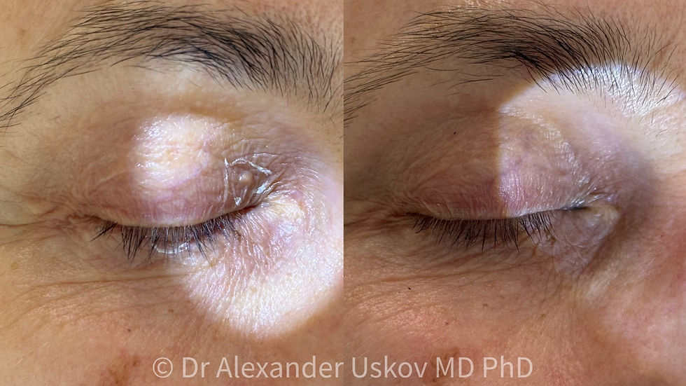
Clinical example from Dr Uskov Kliinik: several skin lesions on and around the upper and lower eyelids are marked before removal on the left; the same area after treatment is shown on the right, with the eye closed in both photographs. Photograph © Dr Alexander Uskov, MD, PhD. Individual result; outcomes vary.

Clinical example from Dr Uskov Kliinik: arrows on the left mark skin growths near the inner eye corner and below the outer part of the lash line before removal; the same areas after treatment are shown on the right. Photograph © Dr Alexander Uskov, MD, PhD. Individual result; outcomes vary.

Clinical example from Dr Uskov Kliinik: the white arrow on the left marks a raised skin growth near the outer corner of the eye before removal; the same area after treatment is shown on the right. Photograph © Dr Alexander Uskov, MD, PhD. Individual result; outcomes vary.
What causes eyelid papillomas? Are they linked to HPV?
Not every eyelid papilloma is a viral wart. Common warts are caused by HPV, while the name “papilloma” is also used for non-viral growths. An eyelid bump alone is not evidence of a sexually transmitted infection. [1]
Conjunctival papillomas have a stronger association with HPV, particularly types 6 and 11. This is one reason to distinguish an external skin lesion from a growth on the inner eyelid or eye surface. [2]
Papilloma, skin tag, syringoma or xanthelasma?
Several conditions can produce bumps around the eyes. These typical features are useful for understanding the differences, but overlap is common. [4,9]
Growth | Typical appearance |
|---|---|
Squamous papilloma or skin tag | A soft, raised growth, sometimes hanging from a stalk. The terms can overlap. |
Viral wart | A rough growth, sometimes with small finger-like projections. |
Syringoma | Small, firm bumps derived from sweat ducts, often clustered symmetrically below both eyes. |
Xanthelasma | Yellowish, relatively flat plaques, often near the inner corners of the eyelids. |
Milium | A tiny, white, dome-shaped cyst. |
Moles, seborrhoeic keratoses and some skin cancers can also resemble a harmless growth. When the diagnosis is uncertain, the doctor may recommend a biopsy rather than cosmetic removal. [3]
Eyelid hidrocystoma: a different type of lump
A hidrocystoma is a benign cystic growth arising from sweat glands. It is a different type of eyelid lesion from a papilloma. [10]

Clinical example of a lower-eyelid hidrocystoma before removal (left) and after removal (right). Photograph © Dr Alexander Uskov, MD, PhD. Individual result; outcomes may vary.
Is an eyelid papilloma dangerous? When should it be checked?
A confirmed benign papilloma is usually harmless. The concern is that an unfamiliar eyelid growth may be something else. Arrange prompt assessment if it is enlarging, repeatedly bleeding, ulcerated, changing in appearance, or associated with missing eyelashes or distortion of the eyelid edge. Even a painless, slowly growing lesion can need investigation. [3]
A lesion at the lash line, inner corner or inner eyelid may require an ophthalmologist or oculoplastic specialist. If it affects vision, assessment should not be delayed. [3,5]
How is an eyelid papilloma diagnosed?
The doctor asks when the growth appeared, whether it has changed and whether it causes symptoms. Examination considers the lesion’s surface, its relationship to the lash line and the surrounding eyelid. An ophthalmologist may also examine the inner eyelid and eye surface with a slit lamp. If the appearance is atypical or the diagnosis remains uncertain, a biopsy may be needed to examine the tissue under a microscope. Photographs can help document changes over time, but cannot establish the diagnosis on their own. [1,3,9]
Eyelid papilloma treatment and removal options
Treatment depends on the diagnosis, size and exact location. The choice should account for eyelid function as well as appearance.
Observation
A stable, confirmed benign papilloma can be monitored if it causes no symptoms and does not bother you. Removal is not compulsory simply because a growth is present. [9]
Shave or snip excision
A doctor may remove a suitable superficial lesion under local anaesthesia with a small blade or scissors. This approach can provide tissue for laboratory examination when needed. [5]
Radiofrequency or electrosurgery
These techniques use electrical energy to cut or treat tissue. Radiofrequency excision has been reported for eyelid papillomas and can preserve a specimen for diagnosis. Suitability depends on the lesion and the clinician’s experience. [6]
Cryotherapy
Freezing may be an option for selected lesions, including some viral warts. The eyelid requires particular care because treatment can affect nearby tissue. It should be performed by a clinician, not with a home freezing kit. [3,5]
Laser removal
An ablative laser, such as a CO₂ laser, can remove selected benign eyelid skin lesions. Small studies report encouraging results, but they do not establish that laser is the best choice for every growth or guarantee a scar-free result. Possible complications include pigment changes, scarring and damage to eyelid structures. [7]
Laser ablation destroys tissue, so it may not leave a suitable specimen for histology. If the diagnosis is uncertain or suspicious, biopsy or excision takes priority. [3]
Eye protection is essential during treatment near the eyes. The protection must be appropriate for the laser and treatment location; closed eyelids alone are not adequate. Some procedures require protective corneal shields placed by a trained clinician. [8]
For the clinic’s treatment information and additional clinical examples, see Laser removal of papillomas on the eyelids.
Can you remove an eyelid papilloma at home?
Do not cut, tie off, burn or freeze an eyelid growth yourself. Avoid wart-removing acids and skin-tag removal products near the eye. They can damage skin, cause scarring and delay recognition of a different diagnosis. [5]
What happens after removal?
Aftercare depends on how the lesion was treated. Follow the clinician’s instructions for cleaning and any prescribed products; do not pick a scab or apply unapproved products near the eye. Healing time varies with the size of the wound and the procedure. [5]
Seek urgent medical care if you develop severe eye pain or a change in vision after treatment near the eye. [8]
Ask before treatment about the likely recovery, whether tissue will be examined, possible marks and whether follow-up is needed. A cosmetic result cannot be promised from a before-and-after photograph.
Frequently asked questions
Is an eyelid papilloma contagious?
Not necessarily. A non-viral skin tag is different from an HPV-related wart. The diagnosis determines whether transmission is relevant. [1,2]
Does an eyelid papilloma always need removal?
No. Confirmed benign lesions can be observed. Irritation, visual interference, cosmetic concerns or uncertainty about the diagnosis may change the recommendation. [5,9]
Can it return after treatment?
A growth can recur, and other lesions may develop nearby. Removal of one lesion does not prevent all future eyelid growths. [5,9]
Will removal leave a scar?
Scarring is possible. The risk depends on the lesion, treatment and healing response. A careful technique aims to preserve the eyelid’s contour and function. [7]
Eyelid skin lesion assessment in Tallinn
If you are considering removal, start with an assessment of the growth. At Dr Uskov Kliinik, you can discuss whether laser treatment is appropriate or whether another diagnostic or treatment approach is needed.
This article provides general information and does not establish an individual diagnosis. Clinical photographs show individual results; outcomes and recovery vary.
Sources
Czyz CN, Blair K. Eyelid Papilloma. StatPearls, updated April 2026.
American Academy of Ophthalmology EyeWiki: Conjunctival Papilloma.
Royal Australian College of General Practitioners: Eyelid lesions in general practice.
American Academy of Dermatology: Skin tags—why they develop and how to remove them.
Eshraghi B et al. The use of a radiofrequency unit for excisional biopsy of eyelid papillomas. 2010.
Expert Consensus on Ocular Safety During Laser Procedures. 2025.
NCBI MedGen / National Cancer Institute: Eyelid Hidrocystoma